A fine needle aspirate is one of the most valuable diagnostic tests used when screening for dog cancer.
Key Takeaways
- The cost of a fine needle aspirate is relatively inexpensive considering the value that it adds to the overall examination of a pet.
- Fine needle aspirates allow veterinarians to look at cells to determine if your pet has inflammation, an infection, a cyst, or a mass.
- A very small needle is used to collect cells for a fine needle aspirate. The discomfort is mild and likely similar to what is experienced during a vaccine, injection, or blood draw.
- Although it is possible in very specific cases for a fine needle aspirate to spread cancer, this almost never happens. The possible risk of the test versus the benefit of making a diagnosis should be evaluated.
Fine Needle Aspirate: Simple, Inexpensive, Valuable
If an abnormality, such as a new growth or an enlarged lymph node, is found during an exam, your veterinarian will likely recommend a fine needle aspirate for your dog.
This simple, relatively inexpensive test enables your veterinarian to determine if monitoring is appropriate or if further diagnostics or treatments are needed.
Fine needle aspirates are one of the basic screening tests used when planning to fight cancer. They can help determine if a mass is cancerous. If it is, fine needle aspirates of the neighboring lymph nodes can also tell us if metastasis (cancer spreading) has occurred.
This information is important in formulating the best treatment for pets with cancer.
Because fine needle aspirates collect a small sample size, they may not always provide an accurate assessment. However, this is an extremely useful diagnostic tool for many cancers.
What Happens During a Fine Needle Aspirate
A fine needle aspirate collects a sample from your dog’s tumor or mass by inserting a needle into a target area and removing cells for review under the microscope in a process called “cytology.” Very few supplies are needed to perform this test.
To perform a fine needle aspirate, the veterinarian will take a very small needle and insert it into the area of concern and will either use suction from the syringe to collect cells or will use a “woodpecker” or pecking technique to ensure that enough cells have entered the inside of the needle.
- Sedation is usually unnecessary. Most dogs don’t even feel the tiny needle.
- The veterinarian will then inject the cells within the needle onto a clean microscope slide with an air-filled syringe.
- The sample is gently smeared to create a thin layer of cells for the clinician or pathologist to review (cytology).
- The sample is allowed to air dry and then stained. Staining the slide allows the clinician or pathologist to look for certain characteristics of a particular cell type.
- The cells tell us if there is an infection, inflammation, or possible cancer.
Who Performs Fine Needle Aspirates, and Where
Fine needle aspirates are simple, minimally invasive, and can be performed by general practice veterinarians or board-certified specialists.
If your veterinarian thinks it is needed, they will likely want to do it right away, during your veterinary visit.
Most veterinarians examine the slides themselves and make a report.
Sometimes they also want a pathologist’s opinion. Pathologists specialize in evaluating cells and think about them every day, making them an excellent resource for cases that may be difficult to interpret or diagnose. If a pathologist needs to take a look, your veterinarian will send the sample out to a lab for a pathologist review.
When a Fine Needle Aspiration Is Recommended
Fine needle aspirates can also be useful in evaluating enlarged lymph nodes, masses deep inside the body, organ enlargement, effusions (abnormal fluid within the body), etc.
Fine needle aspiration should be done each time a new growth is found on the body, and sometimes on older growths under monitoring.
It is important to understand that it is impossible to know if a growth on your dog is cancer without a fine needle aspirate. Similarly, your veterinarian can’t be sure it’s benign without a fine needle aspirate.
For example, a common type of skin tumor called a mast cell tumor is cancerous, yet it can feel exactly like a benign fatty tumor called a lipoma.
Assuming that a growth is benign based on how it feels could lead to undetected cancer. No veterinarian, not even an oncologist, can tell if a lump is a cancer just by looking at it or feeling it.
This simple diagnostic test can help rule out cancer, or alert us that we may be facing an ordeal.
Dog Cancers Commonly Screened with Fine Needle Aspirate
Many types of cancers can be screened using fine needle aspirates. Some common types include:
- Lymphoma
- Mast cell tumors
- Melanomas
- Squamous cell carcinomas
- Osteosarcomas
- Sarcomas
Not every tumor aspirates well, as we will note below.
How to Get the Best Results from a Fine Needle Aspirate
Your veterinarian will perform the fine needle aspirate, and because this is often done during the same appointment, no significant preparations are required prior to a fine needle aspirate.
- The procedure can often be performed without sedation for dermal (skin) or peripheral (close to the outside of the body) lymph node aspirates.
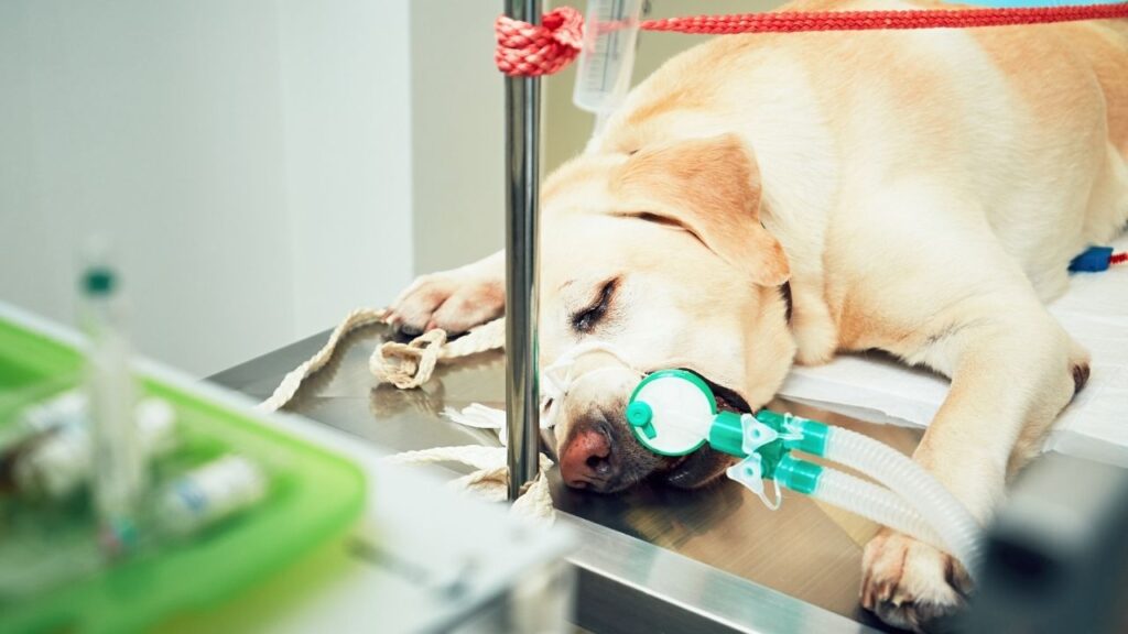
- Sedation may be required or preferred if a pet isn’t tolerant of needle pokes or if the growth is near the eyes, mouth, or a highly vascular area. If a pet is sedated, it is generally preferred for him or her to be fasted prior.
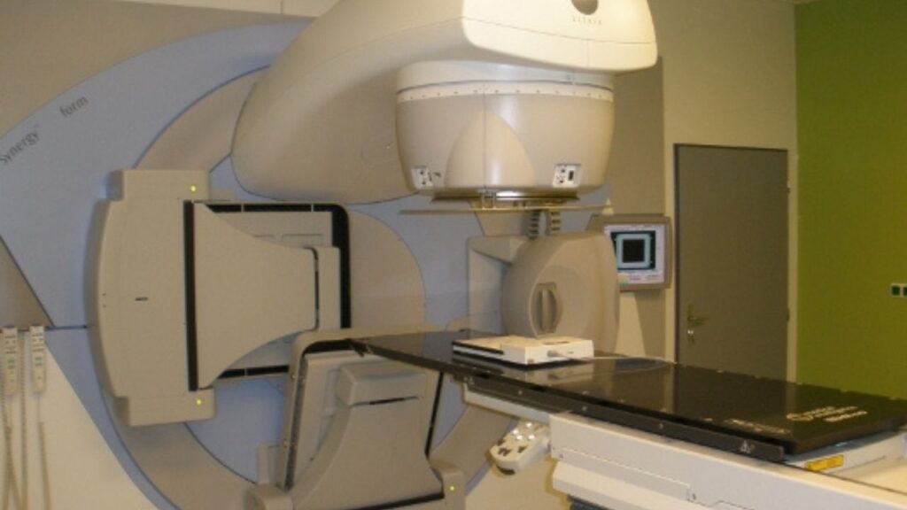
- Aspiration of bones or structures within the chest or abdominal cavity, such as organs, lymph nodes, or masses, requires sedation to get the safest and most accurate results.
Don't worry about lumps. Instead, get them tested, so you know what they are. Early detection pays off, according to veterinary oncologist Dr. Sue Ettinger.
Home Care After Fine Needle Aspirate
Generally, no follow-up care is required at home after a fine needle aspirate. If a pet was sedated for sample collection, it is possible that they will be tired after the procedure, but will likely be completely normal the next day once the sedation medication has been metabolized.
Follow Up Tests for Fine Needle Aspirate
If a fine needle aspirate confirms that a growth is benign, your veterinarian will help you understand your choices for the next steps.
If it’s a cyst or lipoma (benign fatty tumor), it is reasonable just to monitor them, and only treat then if they are growing, changing, causing pain, or upsetting the owner or dog. There are a few exceptions to this, however:
- It’s important to know that cysts have the potential to rupture or get infected at any time. Removing them up front may avoid a more serious problem later on.
- The sizes of lipomas are highly variable and, in some cases, can be quite large. Removing these before growth occurs that impinges on surrounding tissues is preferred.
If the fine needle aspirate confirms a type of infection, appropriate medications, such as an antibiotic, will be recommended.
Finally, if the fine needle aspirate confirms that there are suspicious cells for cancer, a plan will be made to determine the next step for diagnosis and treatment. Since performing a fine needle aspirate is a screening test, follow-up care will be determined by what is found on the sample.
Veterinary oncologist Dr. Brooke Britton explains how cancer is staged and graded on DOG CANCER ANSWERS.
When to Not Use Fine Needle Aspirates in Dogs
Collecting a fine needle aspirate with cytology is an excellent screening tool in veterinary oncology, but it does have some limitations.
- There are specific tumor types that don’t aspirate well, and samples are sometimes non-diagnostic.
- In some cases, the lack of cells on a slide can raise the suspicion of certain cancer types to the veterinarian.
- He or she may recommend obtaining a larger sample via biopsy instead.
Sarcomas and Mammary Masses
A common tumor type that doesn’t aspirate well is sarcomas. Your vet may say that your dog’s tumor isn’t “exfoliating well,” which means it doesn’t shed cells well. It may be too hard for the fine needle to “suck up” cells from these dense tumors.
Mammary masses in dogs are also notorious for fine needle aspirates that are less clear-cut. But that doesn’t mean they aren’t useful.
- We know that fine needle aspirates of the mammary glands in dogs do not correlate well with biopsy results or for prognostication purposes.
- One could argue, however, that fine needle aspirates of growths within mammary tissue could be a good screening tool to look for other tumor types within or adjacent to the tissue.
- One specific tumor type that can occur near a mammary gland is a mast cell tumor, but so can carcinomas. A fine needle aspirate can help your veterinarian determine what type of tumor is present, which helps with choosing treatment plans.
- Larger surgical margins are recommended when removing mast cell tumors, so knowing its presence beforehand would be helpful for the surgeon.
Bladder and Prostate Cancer
Finally, there is controversy about whether aspirating cells from a mass within the urinary bladder or the prostate is safe.
The argument states that there is a risk of spreading cancer cells along the needle track from the inside of the bladder or prostate to the abdominal cavity. Before deciding whether to aspirate a growth from the urinary tract, a pros and cons discussion is needed.
In these cases, a helpful urine screening test called a BRAF test to evaluate for bladder and prostate cancer might also be helpful. The BRAF test may even replace a fine needle aspirate for screening those cancer types.
Safety and Side Effects
- Fine needle aspirates are very safe, and side effects should not be expected under normal circumstances.
- If an aspirate is done in a highly vascular location, the risk of bleeding is possible, but rare.
- Local histamine release (redness, hives, anaphylactic reaction, etc.) is possible if a mast cell tumor is aspirated, although very uncommon.
Fine Needle Aspiration Costs
A fine needle aspirate cost is relatively inexpensive, considering the value it adds to a pet’s overall examination. The price can vary depending on where the test is performed, the lab the samples are submitted to, or if sedation, anesthesia, or imaging is required for acquisition.
Pathologists sometimes charge an additional fee if a special stain is needed to classify a tumor type further.
Fine needle aspirate collection is a screening test, so it is generally not a recurring expense. However, a veterinarian may need to test the site again or order a biopsy if the pathologist requests additional cells. Veterinary clinicians should be able to provide a total cost before the test is performed.
Topics
Did You Find This Helpful? Share It with Your Pack!
Use the buttons to share what you learned on social media, download a PDF, print this out, or email it to your veterinarian.

Editor's Picks


RELATED ARTICLES
ADVERTISEMENT