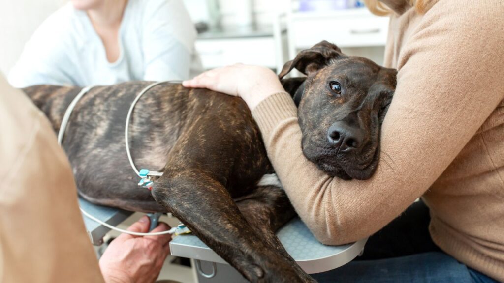
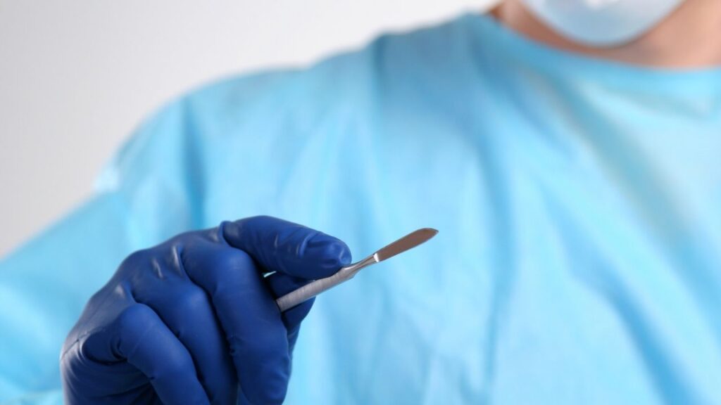
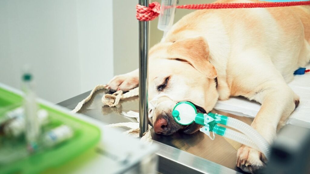
Margins are the areas around a surgically removed tumor. Pathologists examine margins through a microscope to determine if the cancer has been fully removed.
Key Takeaways
- Clear margins mean that there were no cancer cells seen at the edges of a removed tumor. This means that there is a good chance that your dog is now cancer free. However, there is a small chance that a few cancer cells could extend beyond the edges because it is impossible to examine the entire margin.
- Clean margins in dogs mean the same thing as clear margins: there is a good chance that all of the cancer was removed.
- Cancer cells in a margin mean that cancer cells were seen in the marked edges of a tumor. This means that there might still be cancer cells left in the body after surgery. Any cancer cells left in the body can continue to grow into a new tumor or spread to other areas.
- You may see the term “dirty margins” if cancer cells were seen at the edges of a removed tumor.
Knowing the Status of Tumor Margins Helps with Cancer Prognosis
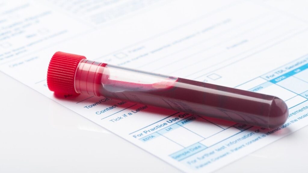
Margins refer to the edges of a tumor resected or removed during surgery and submitted for a biopsy. Pathologists determine margins by examining dyed slices of the tumor under the microscope.
Margin evaluation is one of the most important tools that oncologists use to determine how best to manage their patients to give them the best chance at a good prognosis.1,2
When Margins are Examined
Microscopic examination of tumor margins only needs to be performed if the goal of surgery is to remove the cancer completely.
Margins are typically not examined if the surgery was only done to reduce the tumor size (debulk) to help improve patient comfort.3
Positive/Clean or Negative/Dirty
The pathologist examines parts of (not all of) the margins of the tumor sample. They hope to see a wide section of normal tissue surrounding the tumor sample.
They measure the margins to see how wide they are. They also look for cancer cells in the margins. In their report, they describe the margins as negative (often called “clean”), or positive (often called “dirty”).1,2,4
- Margins are negative or clean when there are no cancer cells found at the edge of the removed tissue and the margins are wide. This means there is a good chance that all of the cancer has been removed.
- Positive or involved margins mean that cancer cells extend to or beyond the edges of the removed tissue, or the margins are narrow. This means that cancer cells are likely still present in the body after surgery.
As always in medicine, using the word “positive” means that something is wrong. We will discuss this in more detail in a moment.
How Wide Should Margins Be?
Cancer types may behave differently, so what is considered wide in one type may not be wide in another.
For example, during surgery to remove a mast cell tumor, it is recommended to remove at least 3 cm (about 1.18 in) beyond the edge of the tumor and one fascial plane deep.5,6 (Fascial planes are sheets of connective tissue that can separate different areas of the body, like muscles, from each other.7)
Does this mean every successful mast cell tumor removal features 3 cm margins? No. Smaller, less invasive mast cell tumor surgeries with margins less than 3 cm can be curative, too.
Guidelines are just that: guidelines. They aren’t hard and fast rules that make or break surgeries. Getting a margin of 3 cm on a mast cell tumor does not guarantee a cure, and getting one of less than 3 cm does not mean it wasn’t a good outcome.
Also, different types of cancer may have very different guidelines when it comes to wide margins. This 3 cm recommendation may not apply equally to other forms of cancer.5
Dr. Demian Dressler and veterinary oncologist Dr. Sue Ettinger discuss margins and their importance in treating cancer.
Does the Examination Include Every Part of the Sample?
Examination of margins is a complex process that requires the pathologist to choose the correct areas to examine. We often assume that pathologists somehow look at an entire tumor, but it’s not possible to do that.
- They must slice and stain the tumor sample, carefully tracking each part.
- They are limited by the equipment they have to use, as only part of a tumor slice can fit in their cassettes (more on that below).
- They may not be able to examine every single part of a tumor sample, so they typically choose the parts that are most suspicious for examination.
- Only a small portion of the tumor margins, occasionally 0.1 – 0.01%, can be reviewed microscopically.3
Read this article to find out more about what a biopsy involves.
What Pathologists Are Trying to Determine
The question pathologists are trying to answer is, “Are the margins wide enough to declare this surgery a success?”
To answer “yes” or “no,” they must consider more than just how much normal tissue surrounds the removed tumor. Pathologists consider multiple aspects of the tumor and the tissues in its environment. They also consider the type of cancer itself.3
For example, if there are cancer cells in the lymphatic vessels in the sample, they are less confident it’s a success.
In short, every tumor sample will tell its own story, and whether its margins are wide enough or not will be dependent upon many factors.
One of the reasons biopsy reports take so long to get back is because of the deep and careful consideration each tumor sample is given.
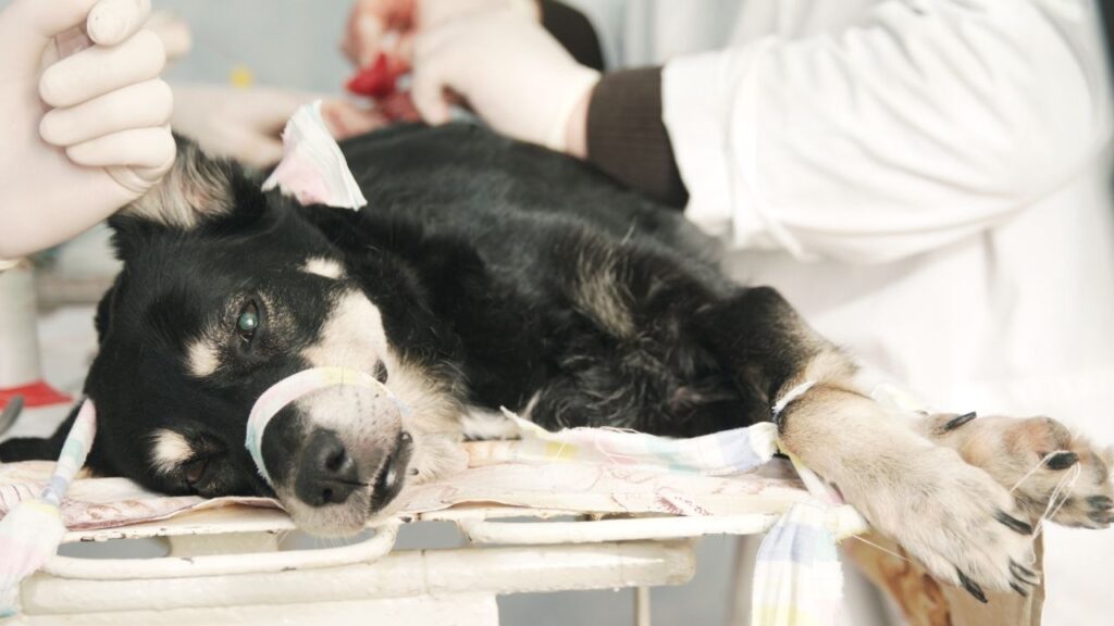
What Happens During a Margin Examination
Dog lovers often wonder why getting the pathologist’s report back takes so long after sending out a biopsy. It usually takes ten days, in part because there are a lot of steps to take to prepare samples before margins can be examined.
- To be examined under the microscope, surgically removed tissue must be cut into very thin slices, around 5-10 microns wide, or the thickness of 1-2 red blood cells.9
- The small width of the slices means there is a limitation on the amount of the tumor and tumor margins that can be realistically examined.
- Pathologists have various methods for cutting and taking slices from the surgically removed tissue to get an adequate representation of the margins.
Different Methods for Different Tumors
In human medicine, tumors are typically cut in a “bread loaf” or “pie” format, which is very labor- and cost-intensive.
We often do not have the same resources in veterinary medicine, so there are some specific ways of sectioning tumors to get the best information about the margins.10
For example, with the “orange peel” method, the outer layer of the tumor is examined, much like an orange peel. These are called tangential sections. They are used to assess the deep and lateral margins for cancer cells extending to the margins.
This method is useful for mammary tumors, feline adenocarcinomas, and mast cell tumors.
The orange peel method is not as helpful for soft tissue sarcomas because they can have tentacle-like projections that are easy to miss. (Dogs with soft tissue sarcomas should have a CT or MRI performed so that surgeons can better know how best to move the tumor.10)
Preparing Samples for Examination
A great deal of time is spent just preparing the tumor sample to be viewed under a microscope. Many specific methods might be used, but a general workflow looks something like this:
- The tissue is dyed, sometimes with multiple colors.
- The sample is dried and then fixed so the dye doesn’t migrate.
- The fixed sample is cut and sliced very carefully, and then pieces are inserted into little cassettes.
- Held in these cassettes, each little piece has its water completely removed and then is dipped in hot paraffin so the wax can fully penetrate the tissue.
- The waxed tissue is then cooled down until it is solid. Then, it can be cut into sections.11,12
- These paraffin block sections of your dog’s tumor can be stored for long, even years because the wax preserves them.
The next step is to cut the block to view under the microscope.
- The paraffin blocks to be examined are sectioned into very thin slices so they can be examined under a microscope.11
- Depending on what the pathologist is trying to view in the sample, they may use special stains.
With all these steps that must be taken before a pathologist can even view the samples, it’s no wonder it takes so long to get a biopsy report back from the pathology lab.
Good News: Negative, Not Involved, or Clean
If you see the words “negative,” “not involved,” or “clean” margins on a pathology report, it is usually good news.
Tumor margins are considered clean, negative, or not involved if no cancer cells are seen at the edge of the surgically removed tissue. The pathologist will report the width of the margins they found.
If a margin is clean, there is a good chance that all the cancer was removed, no additional surgeries are needed, and the risk of the cancer regrowing is low.
But celebration might be a little guarded: remember, it is nearly impossible to examine the full margin of a tumor, so there may be a small number of cancer cells at the margin in an area that was not examined.
This possibility is a reason wide surgical margins are attempted during surgery. Surgeons want to improve the odds of getting clean margins.1,4,5
Bad News: Positive, Involved, or Dirty
If you see the words “positive,” “involved,” or “dirty” margins on a pathology report, it is usually not good news.
Dirty margins are also known as positive or involved margins. You will see these words when the pathologist has noticed cancer cells reach the edge of the surgically removed tissue.
Dirty margins mean that there are likely cancer cells still in the body that have not yet been removed. This means a second surgery may be needed to prevent the cancer from spreading back or spreading.1,4
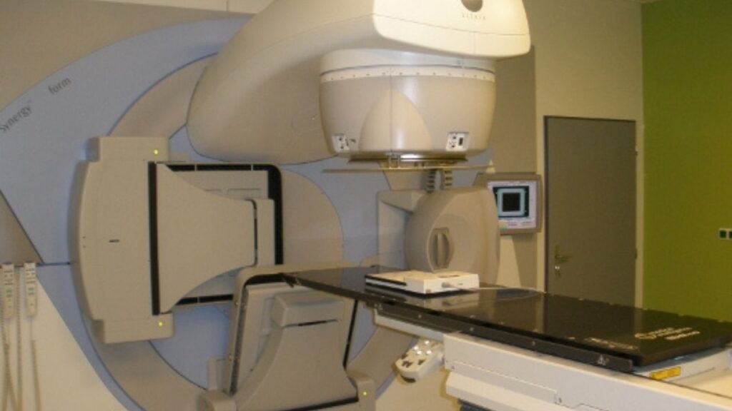
It is difficult to get clean margins when tumors are in areas near blood vessels, nerves, or against the body wall. In these cases, radiation and chemotherapy might be recommended to reduce the growth of the remaining cancer cells.14
Wide Margins
The amount of tissue removed during surgery is critical to lower the risk of dirty margins. Your pathology report might note that wide margins were achieved, which is generally good.
Surgery with wide, clean margins suggests a better prognosis for multiple forms of cancer like mast cell tumors, squamous cell carcinomas, sebaceous and apocrine gland tumors, and peripheral nerve sheath tumors.14
What is considered a “wide margin” may vary depending on the tumor type and the location in the body.
Margins wider than the standard recommendation, also called radical margins, may be needed. In these cases, a large amount of healthy tissue must be removed. Radical margins may be needed for highly malignant tumors or where cancer can spread easily to nearby areas.6,14
Narrow Margins
Your pathology report might note narrow margins. Narrow or close margins mean that cancer cells come right up to the margin of the removed tissue but do not cross it.1,4
Narrow margins are often treated as dirty because cancer cells come within as little as 1-5 mm of the margin.15
When cancer cells are located so close to the margin, there might still be cancer cells left in the body from areas the pathologist does not see. As a result, an additional surgery may be needed to create wider clean margins (this is sometimes called “scar revision”). 5
Deep Margins
The pathology report might refer to the deep margin specifically. The deep margin refers to the base of the surgically removed specimen. For example, in a skin tumor, the deep margin would be farthest from the skin’s surface.
Getting a clean, deep margin is always a goal in surgery. However, it is difficult to see, and surgeons often must determine where to cut through touch or palpation. As a result, cancer cells are more likely to be left behind at the deep margin.3
Sometimes, CT or MRI imaging might be performed before the surgery so that surgeons have a more complete understanding of where to make the deep margin.
In one study in human medicine, the deep margin was involved in 87% of tumors with dirty margins. This study shows how important getting a clean, deep margin can be.5,16 It is very important to have the deep margin marked by the surgeon so that a pathologist can ink it and look there for any cancer cells that might be left in the animal.5
Dr. Demian Dressler and veterinary oncologist Dr. Sue Ettinger discuss when tumors are good candidates for surgery.
Costs
Some commercial laboratories do not automatically perform margin evaluations with their biopsies. Your veterinarian will check to ensure they can perform the service before sending the sample.
Many of the top facilities for margin evaluation are located in diagnostic laboratories associated with veterinary schools.
The cost for obtaining a biopsy and margin evaluation will vary based on the facility, whether they use a commercial laboratory or not, as well as the size, number and complexity of the samples to be analyzed.
Margin evaluation requires additional processing steps, and your veterinarian may need to request it separately.10
Margin Evaluations Take Time, But That’s OK
The examination of the biopsy margins dramatically impacts the decisions you make with your veterinarian.
When planning your dog’s surgery, your veterinarian’s goal is to remove the tumor, removing only enough normal tissue to ensure the pathologist can return a reliable diagnosis. This is trickier than it sounds.3
- Some tumors will be in areas where it’s relatively easy to operate.
- Others may be in small areas or very close to critical organs.
- Some cancer types are more likely than others to have tentacles that have branched out. To get adequate margins, these may need even more healthy tissue removed than expected.
- Some tumor types are more challenging to handle than others. A tumor that is solid and encapsulated might almost “pop out,” while another that is squishier might be slippery and messy.
- Other structures in the area, like fascial planes, might prove barriers to adequate margins, especially if the tumor is pressed against them, making the surgery more challenging and the pathologist’s job harder.
You may have noticed that veterinarians, like human physicians, do not like to guess. They do not want to write “clean margins” unless they are very confident in their determination. Patience is necessary while we wait for them to do their work.
In the end, it’s worth the wait. The pathology report will guide you and your team to the treatment plan most likely to help your dog, whether that is another tissue sample or moving on to choosing treatments.
Topics
Did You Find This Helpful? Share It with Your Pack!
Use the buttons to share what you learned on social media, download a PDF, print this out, or email it to your veterinarian.

Editor's Picks


RELATED ARTICLES
ADVERTISEMENT