Fibrosarcoma in dogs can show up literally anywhere in your dog’s body. A type of soft tissue sarcoma, they are slow growing and not prone to metastasize, but they are locally invasive. Early detection and prompt surgery with wide margins is key to a good outcome.
Key Takeaways
- Fibrosarcoma in dogs is locally aggressive, but usually not prone to metastasis. Getting wide margins during surgery is important because these tumors can recur, or come back, later.
- Fibrosarcoma isn’t necessarily “curable,” but early and assertive surgery (and follow-up treatments as necessary) lead to better outcomes.
- Dogs with fibrosarcoma aren’t necessarily in pain, although if there is swelling present, you can assume there is discomfort. Oral fibrosarcoma can cause pain, as any mouth tumor can. Fibrosarcoma in the nasal passages can also put pressure on those delicate areas, and cause pain. The rarer bone tumors might also cause pain.
- The costs for fibrosarcoma surgery will vary widely and depend entirely on the surgery that is needed. Smaller tumors may be removed with wide margins in your veterinarian’s office, but larger or more invasive tumors may require a specialist and cost more.
- The prognosis for fibrosarcoma depends upon the grade of tumor and whether there is metastasis at diagnosis. You may be looking at months, for high-grade tumors, to years for low-grade tumors that are quickly and entirely removed.
Fibrosarcoma in Dogs: a Type of Soft Tissue Sarcoma
Fibrosarcoma (also called by its acronym, FSA) is a type of soft tissue sarcoma.7 These malignant tumors can arise in many places in the body, because they occur in a type of cell called a fibroblast.1 Fibroblasts are very commonly found in soft tissues throughout the body.
“Soft tissues” in medical terms include muscle, fat, blood vessels, the fibrous tissue found in tendons and ligaments and fascia, and other tissues that provide support to the body.
Fibroblasts are widespread in soft tissues. They connect tissues, organs, and bones within the body and provide structural support.11
No Matter Where It Is Located, a Fibrosarcoma in Dogs Develops in Fibroblasts
Because of the role and location of fibroblasts throughout the body, fibrosarcoma most often occurs in and beneath the skin, on the limbs and trunk of the body.
However, a fibrosarcoma can also take root in the nasal cavity and mouth, as well as in bones.10
If left untreated, a fibrosarcoma tumor will continue to grow, though typically slowly (unless the tumor is of a high grade, or more aggressive).
Stats and Facts About Fibrosarcomas in Dogs
- Fibrosarcoma is the third most common type of malignant oral abnormal growth in dogs, with a prevalence range of 8% to 25%.5
- Fibrosarcoma is known for its ability to reoccur. Studies have found it comes back in the same location 32-57% of the time.3, 13
- Fibrosarcoma is not as likely to metastasize as some cancers are: 15% of dogs developed metastases in the lungs or lymph nodes.13
What Causes Fibrosarcomas to Develop?
Very few tumors and cancers have a single known cause, and the reason a particular dog may develop fibrosarcoma is not straightforward. Cancer in general is a “multifactorial” disease, which means several things need to go wrong for it to take root and grow.
Risk Factors for Dogs and Fibrosarcoma
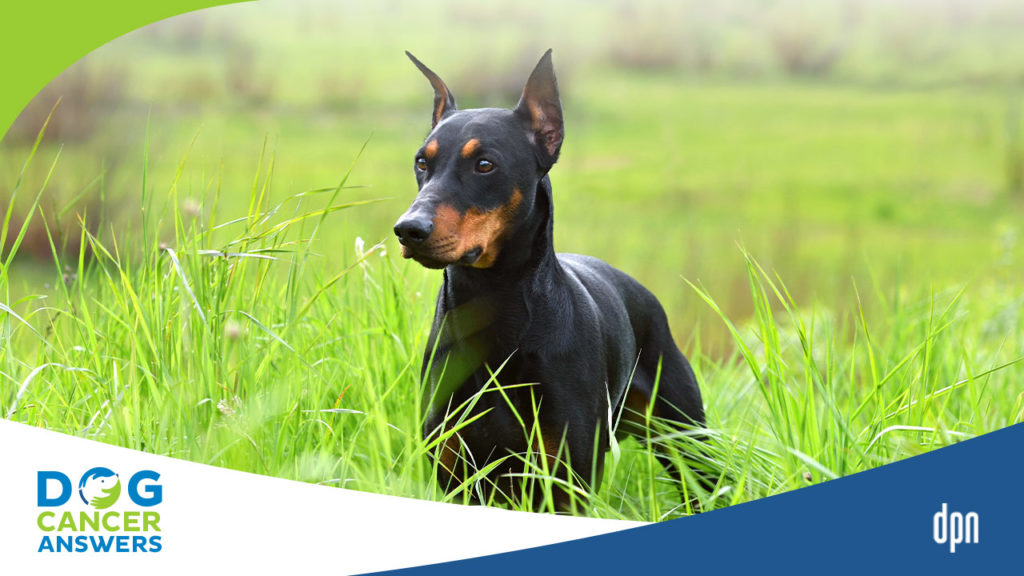
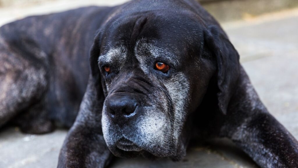
Any age or breed of dog may develop fibrosarcoma, but older and larger dogs are more at risk of developing them (as is true of developing cancer in general). Certain breeds may be at higher risk for fibrosarcoma, including Gordon Setters, Irish Wolfhounds, Brittany Spaniels, Golden Retrievers, and Doberman Pinschers.7
One risk factor increasingly noted is receiving an injection, such as a vaccine or microchip. The development of fibrosarcoma in cats has long been associated with vaccine injection sites, and there is evidence that this may also be the case with dogs.15 (More on this below.)
An amazing True Tail about Tesoro, a Bernese Mountain Dog who fought fibrosarcoma. A must-listen episode of DOG CANCER ANSWERS.
Symptoms of Fibrosarcoma in Dogs
There are many signs you may see in your dog that prompt you to go to the veterinarian.
- Some of these are non-specific, meaning that they could be caused by more than one illness or condition.
- Pain
- Loss of appetite
- Weight loss
- If there is a leg tumor, there might be lameness, difficulty getting up and down, or inability to walk
- A bone fracture with no obvious cause
- If there is a nasal tumor, you may see nasal discharge, nosebleeds, or sneezing
- Oral fibrosarcoma in dogs may cause difficulty picking up food, chewing, or swallowing, and increase in drooling, bleeding from the mouth, bad breath, or loose and lost teeth
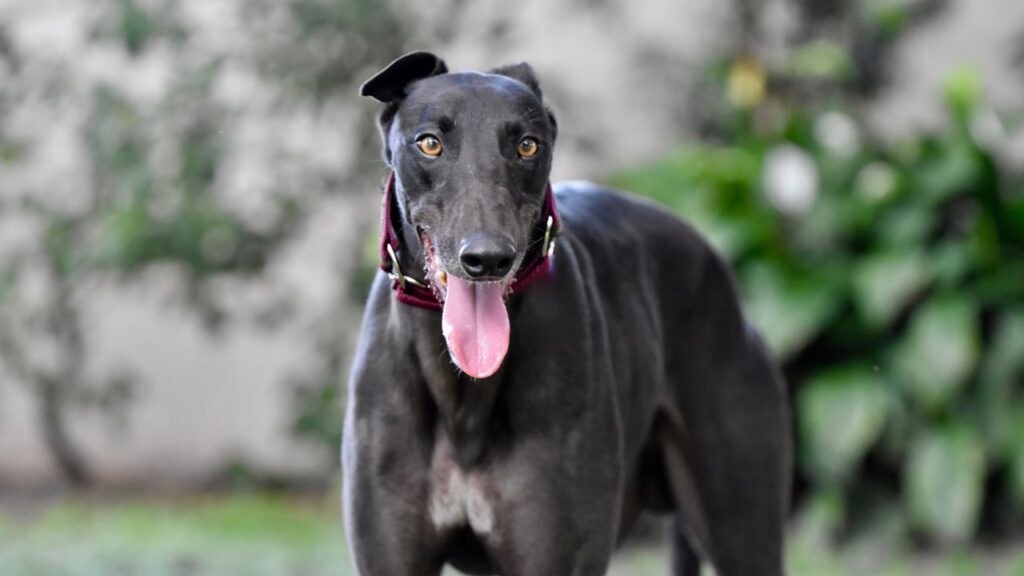
What Fibrosarcoma in Dogs Looks Like
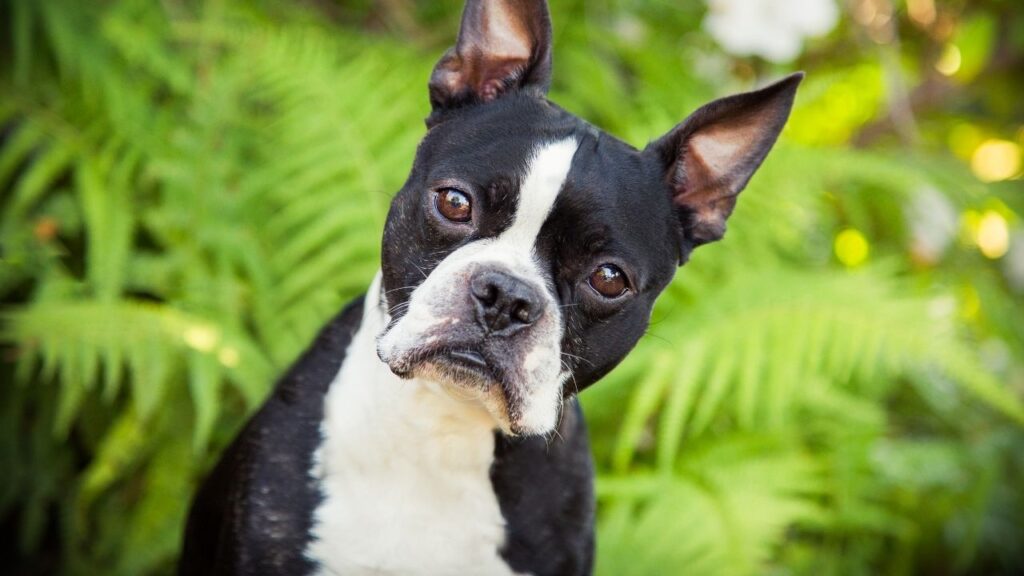
No one can tell if a lump or bump is a malignant cancer just by looking at it or feeling it, and fibrosarcoma vary in appearance and size.
However, since most fibrosarcomas are in the skin or in the tissues just underneath the skin, a fibrosarcoma tumor typically appears as a single, sometimes nodular, firm lump or bump on or under the skin.7
Other fibrosarcomas that occur in deeper areas of your dog’s body might not be seen by the naked eye or felt by the fingers but will appear in imaging.
How Veterinarians Diagnose Fibrosarcoma in Dogs
To get a diagnosis of fibrosarcoma, your vet will need to collect a sample of tumor cells to view under the microscope. This will help them to make a diagnosis and determine how the tumor may behave moving forward.
These tumor cells can sometimes be obtained by aspiration, but more commonly, via biopsy.
Fine Needle Aspirate Is Sometimes Used for Fibrosarcomas in Dogs
Sometimes veterinarians will try using a fine needle aspiration. This is a quick and very minor procedure.
A small needle is used to suction a sample of cells directly from the tumor. These cells are then placed on a slide so that they can be viewed underneath a microscope.
This is a common test used to investigate suspicious tumors. However, this method is not ideal for fibrosarcoma because fibrosarcoma tumor cells stick to one another, making it difficult to obtain a sample.
If your veterinarian is confident that the lump or bump is fibrosarcoma, then they will likely perform a biopsy instead.
Biopsy Is More Often Used for Fibrosarcoma in Dogs
A biopsy is more likely to help your veterinarian come to a fibrosarcoma diagnosis than a fine needle aspirate. This is often a minor surgery, depending upon the size of the tumor your dog has.
During a biopsy, your veterinarian will perform surgery to excise (remove) the tumor. They might take just a piece of the tumor or try to take out the entire tumor itself. The excised tumor tissue is then sent to a lab and examined by a pathologist under a microscope.
Immunohistochemistry
Even when viewing a biopsy under a microscope, fibrosarcomas can be very difficult to discern from other types of tumors. Getting the tumor type correct is important, because your veterinarian might use different treatments for other tumor types.
Because of this, your veterinarian or oncologist may also recommend immunohistochemistry (IHC). In this process, the pathologist uses special stains on the sample. Depending upon what they see, they can differentiate between specific tumor types.8 Pathologists may look for the mitotic index, Ki-67 levels, and how much necrotic (dead) tissue is in the sample.7
Staging for Fibrosarcoma
When you get your biopsy report back from the veterinary pathologist, you will likely see you dog’s fibrosarcoma classified as “low”, “intermediate,” or “high” grade.
- A “low” to “intermediate” grade fibrosarcoma is typically slow growing and less aggressive, with a lower rate of metastasis (cancer spread).
- A “high” grade fibrosarcoma is faster growing and more aggressive, with a higher chance of eventual metastasis.
Once you know what grade your dog’s fibrosarcoma is, you may want to do further staging to determine if it has spread to other parts of the body. Your vet may recommend:
- Bloodwork (CBC, chemistry)
- Urinalysis
- Radiographs (X-rays)
- Abdominal ultrasound
- CT Scan
Surgery is the first treatment choice for fibrosarcoma, and imaging tests like X-rays, ultrasound, and CT scan may be necessary to help your veterinarian plan and execute a successful surgery.
Staging can get quite expensive, so this is an area where you can weigh the pros and cons.
- The pros of staging are that you and your dog’s veterinary team will have a complete picture of your dog’s health and what parts of his body are affected by the cancer. This information can give you a more accurate idea of how he might do and might also rule in or out some treatment options.
- The cons are that it is expensive and does not always give information that will change what you decide to do.
A detailed discussion of the potential benefits of staging for your dog’s specific case should be had with your dog’s oncologist.
With lower rates of metastasis for low and intermediate-grade fibrosarcoma, for example, if budget is a concern, skipping some staging tests in favor of treatment might be a good idea. Oncologists are often the best ones to advise you about whether staging tests are critical or “nice to have.”
Fibrosarcoma in Dogs Life Expectancy
The life expectancy for dogs with fibrosarcoma can really vary widely. Significant predictors of median survival time in dogs with fibrosarcoma include:
- Tumor location
- Tumor size
- Type of surgery performed (aggressive surgery yields longer survival times than conservative/incomplete or no surgery)
- How wide the margins are on surgery
- The tumor’s grade4
Prognosis for Fibrosarcoma in Dogs with No Treatment
Without any treatment, fibrosarcoma tumors will continue to grow and invade underlying tissues. In the case of high-grade sarcomas, distant metastasis (typically to the lungs) is possible.
Prognosis for Fibrosarcoma in Dogs with Treatment
Prognosis is highly variable depending upon the size, tumor location, whether treatment is pursued, and whether there is metastasis at the time of diagnosis.
Median survival times can range from months (in the case of high-grade aggressive or metastatic sarcomas) to years in the case of completely excised, low-grade sarcomas.
Hope for Dogs with Fibrosarcoma
Remember, fibrosarcoma is slow growing overall. This means that favorable outcomes are possible, even if treatment is not curative.
For example, only about 10% of low- to intermediate-grade fibrosarcoma tumors in dogs will metastasize.7
For oral fibrosarcoma tumors, the rate of metastasis is relatively low, which is good news. The less good news is that local invasion into the underlying bone is frequently documented, and complete removal of these oral tumors is challenging.5
Common Treatments for Fibrosarcoma in Dogs
Generally, the focus of cancer treatment in dogs is on improving and maintaining quality of life while also hopefully extending the dog’s life beyond the expected outcome if the cancer wasn’t treated.
The first course of treatment for fibrosarcomas in dogs is always surgery. If you can just cut out the tumor, you may buy good quality time with your dog.
However, fibrosarcoma tumors can occur in inaccessible parts of the body, making full eradication difficult to achieve. Also, because these tumors can recur, even initially successful treatments might feel like failures later on. Once regrowth is observed, there may be a limit to the approach and type of treatments that are offered.14
Don’t lose hope, though. Even if a “cure” is not possible, treatment can greatly enhance quality of life for the time a dog has remaining.
Surgery: Treatment of Choice for Fibrosarcoma in Dogs
Surgery is the treatment of choice, and wide margins are always preferred. Unfortunately, complete removal of the fibrosarcoma may not be possible because so many of these tumors infiltrate surrounding tissues.7
- Large tumors, and those located in parts of the body like the head which don’t give the surgeon much room to work with, are the most challenging cases.18
- A follow up treatment (a second surgery, radiation, or chemotherapy) may be needed if the tumor was not completely removed with the first surgery.
Be prepared for your veterinarian to suggest amputation if the tumor can be removed by removing the body part it is impacting.
- If the tumor is found in a leg, your vet may suggest amputating the limb.
- For oral fibrosarcoma, your vet may suggest a mandibulectomy (complete or partial removal of the lower jaw) or maxillectomy (removal of the upper jaw and palate) to improve outcomes.18 For dogs with oral fibrosarcoma in which surgical excision was the sole treatment, investigators reported median survival times of 7 to 33.7 months and 1-year survival rates of 21% to 50%.9, 14, 18
In addition to the tumor itself, lymph nodes may also be removed during surgery, if they are near the tumor site, and if metastasis is evident.5 However, this is not very common. In one study involving 40 dogs with oral fibrosarcoma, only 1 had evidence of metastasis to the local lymph node at the time of surgery.13
It is good to keep in mind that surgery can come with complications. Some rare complications reported in studies include:
- Major hemorrhaging or blood loss, possibly requiring a blood transfusion.
- Dehiscence (reopening of the surgical incision).
- Infection
The cost of surgery to remove a fibrosarcoma will vary depending on how big the tumor is and its exact location in the body. Smaller, simpler surgeries may be done by your general practice veterinarian. More complex and specialized surgeries should be done by a specialist surgeon and will likely cost more.
Chemotherapy: a Possible Follow-Up Treatment for Fibrosarcoma in Dogs
Chemotherapy is generally not used as a primary course of treatment for fibrosarcoma because it is more helpful for metastasis or systemic cancers. In some cases, chemotherapy might be recommended as a follow-up treatment to surgery, or when surgery is not an option.
Radiation: A Clean-Up Treatment after Surgery
Radiation is not usually a primary treatment for fibrosarcoma, because when used alone without surgery, it’s associated with poorer outcomes.13 That said, it may be suggested for oral fibrosarcoma in dogs for whom surgery is not possible, due to tumor size or location, or if you as the owner do not want your dog to undergo surgery.13
Most often, radiation is suggested as a treatment to be used in conjunction with or as a follow-up to surgery.
- In one study of 28 dogs who received surgery and radiation, a median survival time of 505 days was reported.3
- In another study of 8 dogs, a median survival time of 576 days was reported. This study also found that a combination of radiation and surgery was the strongest predictor of prolonged survival.4
The primary complication associated with radiation pertains to dogs with oral or nasal fibrosarcoma. Radiation treatment in the mouth and nose can cause:
- Oronasal fistulae (an opening between the oral and nasal cavity)
- Mucositis (inflammation of the oral cavity)
- Mouth ulcers
- Discomfort
In a study of 17 dogs who underwent radiotherapy for oral fibrosarcoma, two developed oronasal fistulae. One of these dogs tolerated the condition but the other had to be euthanized due to complications.13
Immunotherapy
Currently there are no immunotherapy treatments for fibrosarcoma in dogs.
Vaccine
There is no vaccine used in the treatment of fibrosarcoma, but the injection sites of previous vaccinations have been associated with the development of fibrosarcomas.16 (See more on this below.)
Diet for Dogs with Fibrosarcoma
There are no specific dietary suggestions targeted for fibrosarcoma. The most important thing when feeding a dog with fibrosarcoma is to make sure that her diet is complete and balanced, providing all of the nutrients that her body requires. Commercial foods should have an AAFCO Statement on the packaging guaranteeing the nutritional value of the food, and home-made diets should ideally use recipes formulated by a veterinary nutritionist.
Supplements for Dogs with Fibrosarcoma
There are no supplements specifically targeted for fibrosarcoma, but there are some on the market for cancer as a whole. Always consult your veterinarian before adding a supplement to make sure that it is a good fit for your dog and that it won’t interfere with any of her medications or treatments (for example, artemisinin should not be given during radiation therapy). Also only add one supplement at a time, so that if your dog reacts to it poorly, you will know exactly which thing she didn’t tolerate. In most cases it is better to choose a few supplements that target your dog’s primary needs, rather than throwing a bunch of supplements at her. This will also help to keep costs down.
Integrative Therapies
No integrative therapies specific for fibrosarcoma are known, although therapies like massage, acupuncture, and rehabilitation may help with your dog’s wellbeing, pain management, and life quality.
What the End of Life Looks Like for Dogs with Fibrosarcoma
Fibrosarcoma is typically slow spreading, but will continue to grow if left untreated, or if your dog is not responding to treatment.
In advanced cancers, metastasis does sometimes occur. Other problems associated with fibrosarcoma in dogs at the end of life are:
- Tumors ulcerate and keep bleeding.
- A tumor recurs and lowers life quality.
- Oral fibrosarcoma tumors may cause difficulty in eating.
- Nasal tumors might make breathing difficult.
If at any point you are concerned about your dog’s life quality, a discussion with your veterinarian about hospice care might be reassuring and meaningful. If your dog’s quality of life slips enough, you may need to face the difficult choice of humane euthanasia for your dog. Always trust your gut when it comes to end-of-life decisions. You know your dog best, and you understand what constitutes quality of life for them and for you.
Prevention Strategies for Fibrosarcoma in Dogs
It is difficult to pinpoint specific preventative tools for fibrosarcoma because concrete causes are unknown. Many factors that can lead to cancer arising (like genetics) simply cannot be controlled. As always, a good diet, exercise, lots of fresh air and good quality water all help to support a healthy dog in general.
Vaccinations, Microchips, and Fibrosarcomas in Dogs
There are several studies linking the development of fibrosarcoma to injection sites; either the sites of vaccine administration or microchip insertion.15,16
Some might conclude that this possible link means you should refuse all vaccinations and microchips for your dog, or for your next one, in hopes that it will prevent fibrosarcoma. But most fibrosarcomas are not directly linked to an injection site. Research into this phenomenon is still in its infancy and a lot more research is required to find the reason for the association, and to troubleshoot what might be happening.
In the meantime, vaccinations and microchips provide many benefits to your dog, and in some localities are legally necessary. The risk of contracting one of the diseases vaccines prevent is higher than the overall risk of developing a fibrosarcoma.
Overall, the risks are far lower than the benefits when it comes to appropriate vaccines and microchips for dogs. Be sure to consult with your veterinarian about which vaccines are important, and which can be skipped before discounting them altogether.
Topics
Did You Find This Helpful? Share It with Your Pack!
Use the buttons to share what you learned on social media, download a PDF, print this out, or email it to your veterinarian.

Editor's Picks
RELATED ARTICLES
ADVERTISEMENT