Ultrasound examination in dogs is a very safe and noninvasive tool that provides detailed pictures and videos of tissues inside of the body. It is most often used to look at organs in the abdomen.
Key Takeaways
- In a dog ultrasound can detect tumors, changes in organ thickness or consistency, fluid pockets, heart function, and pregnancy.
- Ultrasound is performed on a dog by shaving the hair and applying alcohol or gel to the skin to create a liquid interface. The ultrasound head is then gently moved back and forth across the skin to allow the vet to see inside the body.
- A dog should have an ultrasound if your veterinarian is concerned about problems in the abdomen or with the heart, especially if everything looks normal on an x-ray.
- Ideally, dogs should not eat before an abdominal ultrasound. Food in the GI tract can block the view and make it more difficult for your vet to see your dog’s organs.
- Ultrasound is not painful for dogs.
- Ultrasound for dogs typically costs between $300 and $700 but will vary depending on your location and the skill of the practitioner.
- Prepare your dog for an ultrasound by following any instructions from your vet, such as withholding breakfast.
- Dogs have to be shaved for an ultrasound so that the hair doesn’t interfere with image quality.
What Happens in a Dog Ultrasound Examination
Ultrasound is an imaging method that is extremely safe, non-painful, and noninvasive. Dog ultrasound uses sound waves to visualize the structures inside your dog’s body (more on how it works later).
Your veterinarian may offer a quick ultrasound when you bring your dog in after first noticing she isn’t well, but a thorough ultrasound exam is often scheduled in advance. Your veterinarian may recommend scheduling an ultrasound with a boarded radiologist or an internal medicine specialist – these professionals have the most advanced equipment and significant experience reading and interpreting ultrasounds.
An ultrasound may also be done at an emergency clinic to get quick answers about what is going on with your dog.
Abdominal ultrasounds are the most common, but ultrasound can also be performed on the heart, soft tissues like muscles and ligaments, the neck, and other areas. It is useful anytime it is valuable to look under the skin or within a specific body area or cavity.
What to Expect
If your dog’s ultrasound is scheduled in advance, the vet will probably instruct you to fast her (no food, water is ok) for 12 hours prior to the imaging. This is most helpful for abdominal ultrasound so that the most detail can be seen without food obscuring any important areas.
Often the vet will shave the area of interest so that the best images can be taken. Fur is difficult for the machine to see through, so it is necessary in most cases to remove fur in the area that is being imaged.
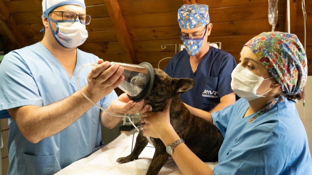
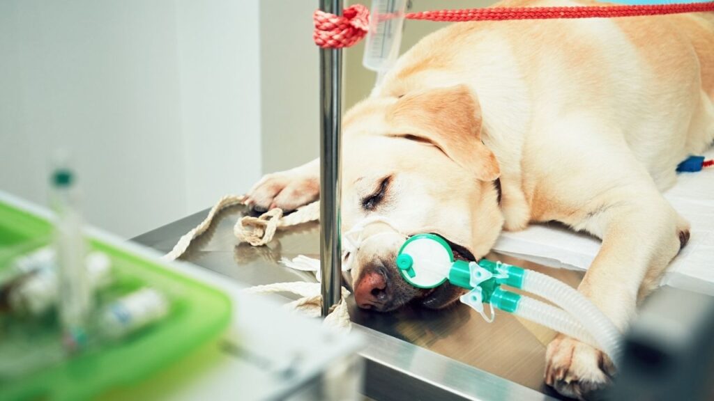
Sometimes, a patient may be sedated or given medications to relax them and make them more comfortable. This reduces stress for the pet and typically results in better pictures on ultrasound because the patient is more still and more relaxed. That said, ultrasound can often be done on a completely awake dog as long as the dog cooperates.
Once the patient’s fur is shaved and they are sedated (if appropriate), special gel or isopropyl alcohol is spread on the skin over the area of interest. This is because ultrasound uses sound waves to create images, and sound travels better through liquid than air. This liquid barrier allows for better penetration of sound waves than is possible over dry skin. Better penetration of sound waves means more accurate and detailed images can be created.
For an abdominal or chest ultrasound, your dog will be placed on her back in a foam trough and rotated periodically to give the vet clear views of the entire area of interest. The ultrasound probe (a small wand-like tool with a flat or slightly curved surface) is moved over the skin with gentle pressure. The probe is attached to a cord that plugs into a computer with a screen that displays the image in real time.
Sound waves go from the probe, through tissue, and back to the probe. This process creates detailed images of tissues, organs, and blood vessels under the skin.
Some machines allow the vet to take pictures or even videos. This is especially helpful if your dog is being seen by multiple vets, because your entire veterinary care team will be able to see the ultrasound images.
Once all images and videos are taken and saved, the alcohol or gel is wiped from the skin and the procedure is complete.
Who Does the Ultrasound Exam?
Your regular general practice vet may have an ultrasound machine and be comfortable using it. It is fairly common for a general practice vet to do a basic ultrasound to confirm any suspicions they have after doing a physical exam plus or minus x-rays but refer patients out to a specialist for a complete ultrasound exam.
For example, if your vet can feel a mass in your dog’s abdomen but it doesn’t show up clearly on an x-ray, she may do an ultrasound to verify that a mass is present and see what it is attached to. If you dog is then referred to a specialist, the specialist will do a more complete ultrasound to get more information about the mass and possibly take samples of it.
If your vet doesn’t have an ultrasound, isn’t experienced reading ultrasounds, or your dog’s ultrasound is difficult to interpret, you will likely be referred to an internal medicine specialist or a board-certified radiologist. These professionals use ultrasound every day, often have more advanced equipment, and are well versed in interpreting complex cases.
Some places have trained technicians do the actual ultrasound and then a radiologist reviews the recording, but usually the radiologist or internal medicine specialist does the actual imaging procedure.
Common Examples of How Ultrasound is Used in Dogs
One of the biggest benefits of ultrasound is that it provides a safe way to look inside the body and create pictures of tissues and organs in real time.
Ultrasound can be used to evaluate:
- Organs like the kidneys, liver, gastrointestinal tract, pancreas, spleen, gall bladder, adrenal glands, bladder, uterus, prostate, lungs, and heart
- Some glandular structures
- Muscle
- Tendons
- Any other soft tissue structure
This technology can measure the thickness of intestinal and stomach walls as well as lymph node size and other real- time measurements.
It can also be used to search for fluid in spaces like the abdomen and chest. This could be blood or other fluids. Sometimes ultrasound is used to guide the process of sampling these fluids with a needle. This allows the doctor to see where the needle is going in order to be most accurate. This aids in extracting fluids and sampling urine from the bladder and can also be used to guide sampling of possible abnormal tissue within an organ.
Doppler
Doppler is a technology used to detect blood flow direction and intensity. Some, but not all, ultrasound machines are capable of this. This technology is useful when evaluating a tumor or suspicious tissue that may be a tumor because the blood flow in tumor tissue is usually abnormal and distinct from healthy tissue.
Doppler can also be helpful in looking at the heart, and can help a doctor avoid important blood vessels when taking fine needle aspirates throughout the body.
When Dog Ultrasound is Recommended
Ultrasound is recommended in many contexts. Its most common use in dog cancer is for searching for potential cancer locations or identifying if and where known cancer has spread.
Additionally, ultrasound is extremely useful for guiding the sampling of tumors or tissues that are known or suspected to be affected by cancer. If a doctor can see inside the body in real time, it is much easier to get an accurate sample of tissue or fluid for diagnostics and analysis.
One common use of ultrasound is to see the bladder when performing a cystocentesis (poking a needle into the bladder and drawing out urine). This procedure is often used to get a urine sample from cats and is also perfect when a sterile sample is needed to do a urine culture and sensitivity.
Another area where ultrasound shines is evaluating heart function in real time. An echocardiogram uses ultrasound to visualize the structure of the heart and to watch how blood flows through the chambers and valves with each heartbeat.
Ultrasound is also used to confirm pregnancy in dogs.
How Dog Ultrasound Works
As mentioned before, ultrasound uses sounds waves to make pictures of tissues in the body.
Different tissues and fluids have different textures, thicknesses, etc. This determines how much of the sound (ultrasound waves) that makes it to the tissue or fluid bounces back to the probe and how much passes through the tissue.
The differences between these tissue properties result in varying shades of white, grey, and black on the ultrasound image. Fluid does not bounce back very much of the sound signal it receives and shows up as black on the screen. Alternatively, tumor tissue and other tissues like organs bounce back more sound waves and show up as varying shades of grey depending on the density and quality of the tissue. Bone and air will also reflect many of the sound waves that hit them.
Ultrasound images are often very difficult for dog lovers to understand, and just look like grey static. But to a trained veterinarian, those swirls and shades of grey reveal organs, blood vessels, bones, and tumors. Many vets talk out loud as they perform an ultrasound to let you know what they are seeing.
Cancers Ultrasound Is Commonly Used For
Ultrasound is used for many different cancer types because of its ability to look inside of the body. Here are some of the cancers that ultrasound can be used to identify:
- GI tract cancers
- Pancreatic cancer
- Kidney cancer
- Heart cancer
- Liver cancer
- Spleen cancer
- Lung cancer
- Bladder cancer
- Uterine cancer
- Gallbladder cancer
- Looking at lymph nodes and measuring their size for signs of metastasis
Ultrasound can pick up abnormalities in tissue texture, size, or quality. While the image itself does not always provide an answer as to whether the abnormality is cancer or what type, often the images can guide testing or sampling of the tissue cells themselves to identify why it looks unusual on an ultrasound image.
Ultrasound can also be useful for monitoring your dog during and after cancer treatment. A quick ultrasound can reveal if your dog’s tumor and/or lymph nodes have shrunk (indicating remission) or increased in size (indicating progression of the cancer).
How to Get the Best Results
Your veterinarian will handle most of the details when performing an ultrasound, but there are some things you can do to prepare your dog and ensure a successful test.
- Follow fasting instructions for abdominal ultrasounds. An abdomen full of food and poop makes it hard to see organs!
- Consider sedation if your dog is anxious or aggressive. This will minimize stress for your dog, and allow the vet to do her job as quickly as possible.
- Expect your dog to be partially shaved for the ultrasound. Vets can sometimes see a little without shaving, but bare skin gives a much clearer picture and will allow the vet to give you better answers about your dog’s health.
Home Care After Dog Ultrasound
There is little home care involved after ultrasound due to its safety and noninvasive nature. However, be aware that some of your dog’s fur will be shaved and they may be a little more sleepy than usual if sedation was used for the procedure.
Follow Up
Follow-up care will vary depending on what is going on with your dog.
Sometimes recheck ultrasounds are used to monitor if a tumor is shrinking or if treatment is effective. Ultrasound may also be used to monitor the amount of fluid in a body cavity and if that fluid contains cancerous cells.
In many cases, follow-up care after an ultrasound done by a specialist will be directed by your oncologist or regular veterinarian. They will review the ultrasound findings and use that information to give you the best treatment options going forward.
When to Not Use Ultrasound
While ultrasound is extremely useful for dog cancer, there are some situations where it isn’t beneficial.
Ultrasound is not used to look at the brain or spinal cord. MRI or CT are usually much better at getting the level of detail needed in these areas.
Also, while ultrasound can determine tissue quality, it is important to remember that it can’t determine microscopic cell type. Ultrasound alone is not able to identify a specific cancer type and give you a definitive diagnosis (I.e., carcinoma, adenocarcinoma, benign vs malignant, sarcoma, etc.). There are also situations in which inflammatory processes and cancerous processes can look similar in certain tissues.
Where to Get a Dog Ultrasound
Ultrasound is becoming increasingly more accessible. Generally, the gold standard is to have an ultrasound done by a board-certified radiologist or internal medicine specialist, but there are general practice doctors who can perform basic versions of the imaging if they have the equipment and have had extra training. Similarly, some oncologists have basic ultrasound skills as well.
There is also a difference between a formal ultrasound study that provides detailed measurements of all components with complete descriptions and the ultrasound method being used to look for fluid or large very visible tumors on an emergency basis. Many ER facilities have some form of the latter. The amount of information and detail provided depends on the skill of the person performing the ultrasound and the quality of the machine.
While formal ultrasound is generally considered a specialty service, some boarded radiologists and internal medicine specialists are mobile and can travel to general practice facilities in their area to provide this imaging service. Ask your veterinarian about the availability of ultrasound in your clinic and whether or not radiologist services are available locally.
Also, some areas may have an ultrasound technician who can perform the procedure and then a radiologist can remotely interpret those images and videos. This is how ultrasound is performed in human medicine and this pattern is emerging in veterinary medicine in certain places as well.
Safety and Side Effects
Ultrasound is extremely safe and there are no true reported side effects. It is also typically painless although sometimes the pressure of the probe can cause mild discomfort in certain patients. However, this is by far the exception and not the rule.
If your dog needs to be sedated for his ultrasound, there are potential side effects from the residual sedation. These can include lethargy, slow breathing rate, or stiffness from lying in an abnormal position for an extended period of time.
How Much is a Dog Ultrasound?
Ultrasound cost varies depending on who is performing it, how many areas are being evaluated, if sampling is taking place (additional cost), and whether or not it is on an emergency basis.
Generally, the cost varies from about $300-$700.1 The highest cost is associated with a boarded radiologist performing an ultrasound on an emergency basis. Recheck ultrasound for the same problem within a short time period is typically provided at a discounted rate (usually between 30%-50%, but this varies).
Topics
Did You Find This Helpful? Share It with Your Pack!
Use the buttons to share what you learned on social media, download a PDF, print this out, or email it to your veterinarian.

Editor's Picks


RELATED ARTICLES
ADVERTISEMENT